Covering up – Head involution in Drosophila embryogenesis
How do complex organisms develop from fertilized egg cells?
This, without doubt, is the question central to all of developmental biology. Much already is known about the underlying genetic machinery involved in determining whether a cell ends up e.g. as a muscle cell or rather as part of the nervous system. When observing the embryonic development of an organism under the microscope, it becomes apparent, however, that apart from these genetic processes a multitude of complex physical rearrangement of cells and tissues have to occur in order for organs to form and for an organism to take on its final form. Think of organs such as the heart or the lung. They are not mere blobs of cells, which could be easily formed by a single cell dividing over and over again. Instead, these organs have a complex geometry. The heart consists of several chambers, valves, etc. Lungs have a branching architecture, with the finest branches ending up in miniscule cavities, so called alveoli, which are responsible for the uptake of oxygen from the air. The development of such organs therefore must involve complex foldings, closures, rearrangements, and movements of large groups of cells. How do cells know where to move? And what processes are providing the physical forces necessary for these movements to occur? These and related questions are studied intensively in research laboratories all over the world. It is, however, still a long way before we will understand the development of the heart and other complex organs. Instead of studying such complicated cases, researchers therefore concentrate on simpler processes that (hopefully!) are easier to understand. After all, if you cannot understand the workings of a Ferrari, maybe it is a good start to look at a bike instead! Of course, not everything you learn about bikes will also hold true for cars. Still, there is ample reason to believe that understanding a simple means of transport with two wheels will also bear significance for our understanding of a more complex one with four. In developmental biology, instead of studying human development, researchers therefore often study less complex organisms, as for example the fruit fly, which is also known by its Latin name Drosophila melanogaster. Fruit flies are comparably easy to keep in the laboratory and many techniques are available for conducting experiments with them.
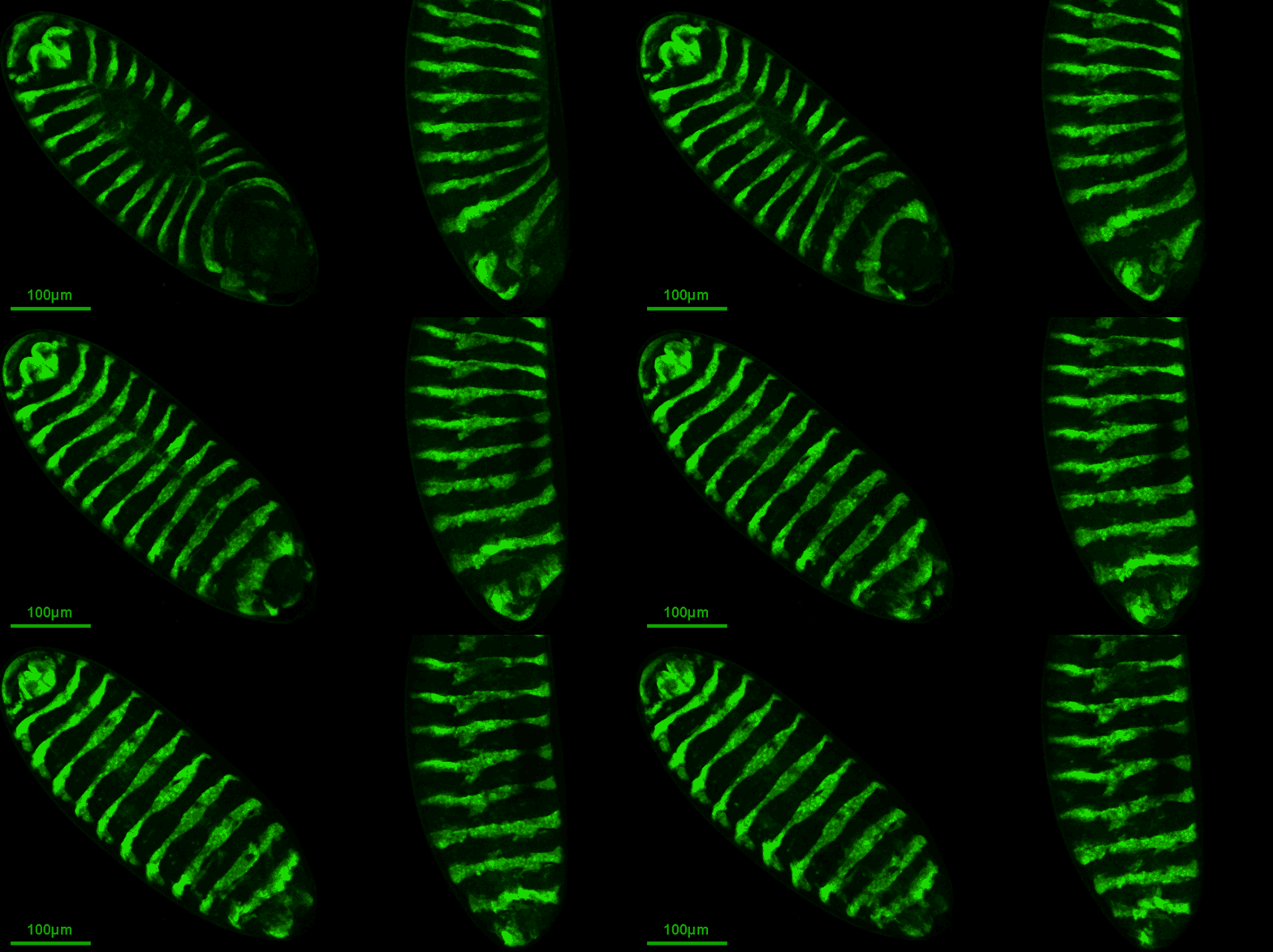
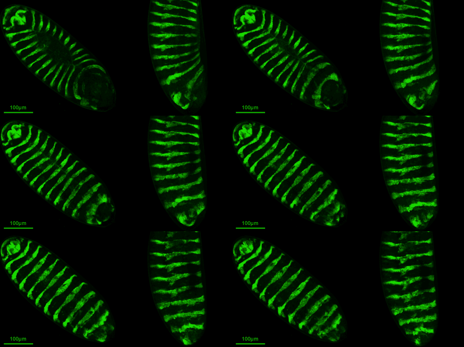
Recently, we have published a paper about a case of tissue movement occuring during the embryonic development of the fruit fly (see movie by Natalia Czerniak at top of post). The development of a fruit fly from a fertilized egg, roughly speaking, has three stages. During the embyonic stage, the fertilized egg develops into a larvae. The larvae then becomes a pupa, just like a butterfly does. And from the pupa, the adult fruit fly emerges. When you think of the larvae, you can picture it as a zeppelin shaped “worm” of roughly half a millimeter length. The inner organs, such as gut, brain, or nervous system, are all enclosed by a continuous sack of skin, also referred to as epidermis. It turns out that during the early phases of its development, this epidermal sack has holes and does not cover the entire embryo at all. In particular, the brain is sticking out at the front side of the embryo! During a process called head involution, then something pretty amazing happens. For one, the brain is pulled backwards, i.e. towards the inside of the forming sack. At the same time, the epidermis moves forward, ultimately covering the brain.
What is fluorescent microscopy?
From own experience, I know that microscopy movies are difficult to understand when seen for the first time. I will try to walk you through it! The movie on top of this post shows the late phases of fruit fly embryonic development. On the left you can see an embryo from the top, on the right one from the side. The head in both cases is at the bottom. You probably wonder: What are the green stripes? To explain this, I need to tell you a bit about how modern microscopes work. Essentially, when viewed through a conventional light microscope, which you can think of as a sophisticated magnifying glass, a fruit fly embryo appears as a little elongated balloon filled with a foggy type of material. Hardly no structure or different parts are distinguishable. Just think of everything having the same colour. Have you ever tried to spot a white rabbit on a field covered with snow? Exactly! In order to make specific parts of the embryo visible, researchers make use of a cool trick. Using modern genetic tools, they modify particular components of the fruit fly, namely proteins, in such a way that they emit fluorescent light when hit by a laser beam. Think of such a component e.g. as a type of screw that helps keeping the embryo together. Instead of the usual screw, the modified fly simply uses a new type of screw, one that lights up when a laser beam is directed at it. Now, one cannot only change all screws in an embryo, but also specific ones. In the case of the movie on top, it is a screw in the epidermis. Not the whole epidermis, however. It turns out the epidermis is striped, and only in each other stripe the screws are changed. Under the microscope, which is attached to a laser, every other stripe thus becomes visible and one can observe the movements of the epidermis over time.
What you can see in the movie are mainly two things. For one you notice a gaping hole when viewing the embryo from the top (left side). This opening closes over time, during a process referred to as dorsal closure. “Dorsal” here is the expert term for “on the back of the embryo”. At the same time, one can see how in the lower half of the embryo one after another the two ends of each stripe are “glued together” to form rings going all around the embryo. Afterwards they start moving towards the front of the embryo (remember, the front is at the bottom here). It is this forward movement which marks head involution. The brain, which is moving inwards, in this movie cannot be seen. Simply cause it was not modified to emit light.
Why does the epidermis move to the front?
This is exactly the question we could answer with our research. Here is what we found! We could show that the epidermis is under tension. Tension is the propensity of a material to contract. Think of an inflated balloon. The elastic rubber of a balloon is stretched when you blow air into it. The material does not like to be stretched, however, and acts against it. This resistance is what you feel when blowing more and more air into the balloon. In the embryo, tension is not generated by air pressure though. Rather it is actively generated by specific molecules in the epidermis, so called motor proteins. These little machines are very similar to the ones at work when contracting a muscle in your body. Now think of the epidermis as made of up rings which are under tension. As you can see, the embryo tapers off towards its end, i.e. it gets smaller. The forward movement of the rings results from the fact that a contracting ring on a tapering object will automatically move towards the end which gets smaller. Have you ever tried to squeeze a piece of soap while being in the bath tub? What does happen in that situation? Exactly, the soap glitches from your hand, simply by you squeezing it! If the soap were fixed, however, it would be your hand that was moving over the soap. And that is exactly why the epidermis is moving over the brain towards the front of the fruit fly embryo! In short, it results from an interplay between the geometry of the embryo and the fact that there is tension in the epidermis. Note, no one needs to tell the epidermis where to move. It moves into the correct direction, simply cause things are set up in the right way!
Of course, in order to show that this is how tissue movements come about in the case of head involution, we had to do many experiments, perform careful analysis of our movies, build computer models, and discuss for quite some months. In a nutshell this is what we found though! If you like to read more, here is a link to a press release that was published on the website of the Centre for Genomic Regulation. This is where the research was performed by my colleagues and myself in the group of Dr. Jérôme Solon.
Also, we were very excited to learn that our publication was recommended by F1000 Prime.
And here is the reference of the original paper:
N. Czerniak*, K. Dierkes*, A. D’Angelo, J. Colombelli, and J. Solon, Patterned contractile forces promote epidermal spreading and regulate segment positioning during Drosophila head involution, Curr. Biol., 2016 (* shared first authors)


Leave a Reply